Shocking Thermal Images from the C19-Injected

Europe Reloaded / Pam Barker

“Computerized Thermographic Imaging and Live Blood Analysis Post C19 Injection”; by Ana Maria Mihalcea, live blood analysis and thermographic Imaging of C19 injected individuals; In these never-before
– seen images of individuals who received the experimental mRNA shot, we can see extensive abnormal vein imaging suggesting blood clotting in both deep venous and superficial venous system.

Summary:
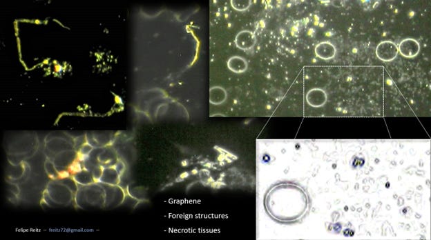
These shocking thermographic images suggest extensive subcutaneous and deep vein blood clotting in C19 injected individuals. Dark Field Microscopy complements the analysis of these individuals, showing what potentially could be Graphene Hydroxide or nanostructures of other metal composition. These alarming findings should alert health care practitioners to screen people who received the gene modifying injections for blood clots, even if they are completely asymptomatic. Thermography may be a noninvasive safe way to do so. Felipe has now also seen this in children as young as 2 years old.
This visual proof of harm should galvanize every health care provider to stop giving the shots and every person to stop accepting them.
See Dr. Mihalcea’s substack below:
****
Computerized Thermographic Imaging and Live Blood Analysis Post C19 Injection
Shocking New Images – Stop The Shots Now!
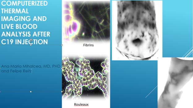
In this interview with Brazilian biologist and medical researcher Felipe Reitz we discuss his findings of live blood analysis and thermographic Imaging of C19 injected individuals. A thermogram is a regional temperature map of the surface of a part of the body, which is obtained by an infrared sensing device. This measures radiant heat and subcutaneous blood flow. Thermography has been proposed as a noninvasive way to detect deep vein thrombosis, which are blood clots in the venous system.
In these never-before-seen images of individuals who received the experimental mRNA shot, we can see extensive abnormal vein imaging suggesting blood clotting in both deep venous and superficial venous system. These individuals are completely asymptomatic and do not feel anything, nor do they have swelling.
Additionally, we are discussing the blood analysis findings of vaccinated individuals compared to unvaccinated.
Watch our presentation and discussion of the shocking findings here:
Computerized Thermographic Imaging and Live Blood Analysis post C19 injection
We correlate the Live Blood Analysis findings with the Thermographic Images.
Recent analysis of C19 shot vials have shown self-assembly nanostructures.
I have written about the results around the world.
Most recently, Mike Adams from Natural News examined the chemical composition of blood clots of injected individuals who died with extensive self-assembly blood clots. The composition showed that this is not the same composition as normal human blood clots, but he found metals like Aluminum, Tin and Sodium that are electrically conductive.
Summary:
These shocking thermographic images suggest extensive subcutaneous and deep vein blood clotting in C19 injected individuals. Dark Field Microscopy complements the analysis of these individuals, showing what potentially could be Graphene Hydroxide or nanostructures of other metal composition. These alarming findings should alert health care practitioners to screen people who received the gene modifying injections for blood clots, even if they are completely asymptomatic. Thermography may be a noninvasive safe way to do so. Felipe has now also seen this in children as young as 2 years old. This visual proof of harm should galvanize every health care provider to stop giving the shots and every person to stop accepting them.
Original Article: https://www.europereloaded.com/shocking-thermal-images-from-the-c19-injected/
Comments ()